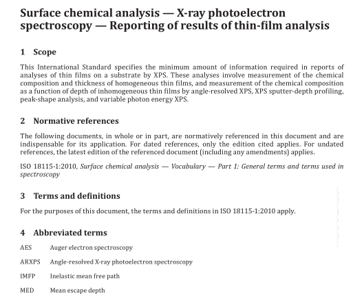
ISO 13424:2013 pdf download.Surface chemical analysis — X-ray photoelectron spectroscopy — Reporting of results of thin-film analysis
5 Overview of thin-film analysis by XPS 5.1 Introduction XPS analyses of thin films on substrate can provide information on the variation of chemical composition with depth and on film thicknesses. Several XPS methods can be used if the total film thickness is less than three times the largest MED for the detected photoelectrons. The MED for particular photoelectrons is a function of the IMFP and the emission angle of the photoelectrons with respect to the surface normal. The IMFP depends on the photoelectron energy and the material. MED values can be obtained from a database. [1] A simple analytical formula for estimating MEDs has been published for emission angles ≤50°. [2] For such emission angles, the MED is less than the product of the IMFP and the cosine of the emission angle by an amount that depends on the strength of the elastic scattering of the photoelectrons in the film. [2] Both the IMFP and the strength versus depend on the chemical composition of the film.
The MED is typically less than 5 nm for many materials and common XPS instruments and measurement conditions. If the effects of elastic scattering are neglected, the MED is given approximately by the product of the IMFP and the cosine of the emission angle. The latter estimates of the MED can be sufficient for emission angles larger than 50° although better estimates can be obtained, e.g. from the database. [1] If the total film thickness is greater than three times the largest MED, XPS can be used under certain conditions (see Annex D) together with ion sputtering to determine the variation of chemical composition with depth. Table 1 provides a summary of the XPS methods which can be used for determining chemical composition and/or film thickness. Some methods can be utilized for the characterization of single-layer or multiple- layer thin films on a substrate and some methods can be used to determine the composition-depth profile of a sample for which the composition is a function of depth measured from the surface (i.e. where there is not necessarily an interface between two or more phases).
The choice of method typically depends on the type of sample and the analyst’s knowledge of the likely or expected morphology of the sample (i.e. whether the sample can consist of a single overlayer film on a flat substrate, multiple films on a flat substrate, or a sample with composition varying continuously with depth), whether the total film thickness is less than or greater than the largest MED for the detected photoelectrons, and the desired information (i.e. film composition or film thickness). The first three methods in Table 1 are non-destructive while the final method is destructive (i.e. the composition of the exposed surface is determined by XPS as the sample is etched by ion bombardment). Brief descriptions of these methods are given in the following clauses and additional information is provided in the indicated annexes.
XPS is typically performed with laboratory instruments that are often equipped with monochromated Al Kα or non-monochromated Al or Mg Kα X-ray sources. For some applications, XPS with X-rays from synchrotron-radiation sources is valuable because the energy of the X-ray exciting the sample can be varied. XPS with Ag X-rays is also used to observe deeper regions compared to excitation with Al X-rays. In some cases, X-ray energies less than the Mg or Al Kα X-ray energies can be selected to gain enhanced surface sensitivity while in other cases, higher energies are chosen to gain greater bulk sensitivity and to avoid artefacts associated with the use of sputter-depth profiling.ISO 13424 pdf download.ISO 13424-2013 pdf download
